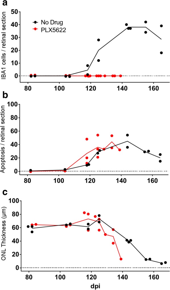
Fig. 4.

Scrapie-induced retinal degeneration in C57BL/10 mice in presence or absence of retinal Iba1-positive cells. On all graphs each dot denotes the mean value of counts from two sections of a single mouse, lines connect mean values at each timepoint, and lines end following euthanasia of final clinical mouse. PLX5622 treatment was initiated at 14dpi. a Number of infiltrating Iba1-positive cells in the photoreceptor layer increased in the no drug (ND) group from 124 to 162 dpi, whereas Iba1-positive cells were not observed in the PLX5622-treated group. b Apoptosis was detected by observation of TUNEL-positive cells in the outer nuclear layer (ONL) in both ND and PLX groups. Apoptosis was not dependent on presence of Iba1-positive cells. c ONL thickness (μm) decreased in both ND and PLX groups indicating that loss of photoreceptor nuclei in the ONL did not depend on Iba1-positive cells. ONL thinning in PLX group which lacked microglia appeared to be slightly faster than in the ND group. However, best-fit line slopes for points from ND and PLX groups between 118 and 164 dpi were not significantly different, p = 0.101
