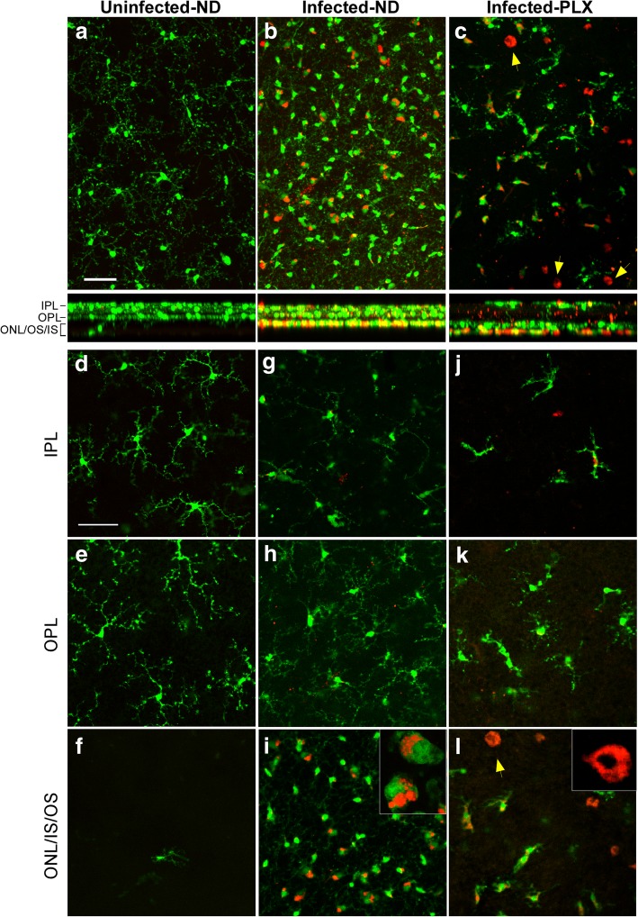
Fig. 7.
Retinal flat mount study of tgGFP/RFP mice by confocal microscopy. a,b,c. Z-stacks of 40–50 optical 1 μm sections of retinal flat mounts from tgGFP/RFP mice which express GFP (green) in microglia and RFP (red) in monocytes. a Uninfected mouse shows scattered green microglia with long delicate processes. Cross-section below with XZ dimension shows most green cells are in the OPL-IPL areas. b At 159dpi infected mouse from ND group has a large increase in green microglia with larger cell bodies and stubby processes typical of activated microglia. Some green/red dual stained cells can also be seen. XZ cross-section shows many of these cells are now in the ONL/IS/OS region. c At 134 dpi an infected mouse from the PLX group shows reduced number of green microglia and a few green/red cells as well. In addition, a few round all red cells, probably monocytes can be noted (arrows). d-f Single optical sections from uninfected mouse show that microglia are mostly in the IPL and OPL layers. g-i Optical sections of 159 dpi ND mouse show that green microglia in the IPL and OPL layers have processes similar to those seen in uninfected mice, whereas in ONL,IS,OS region green microglia and green/red microglia appear activated and have stubby processes. Some cells have red material apparently internalized in phagolysosomes (inset). j-l. Optical sections of 134 dpi PLX mouse also have a few green microglia with processes in IPL and OPL layers. In ONL/IS/OS region round cells with red cytoplasm appear to be monocytes (arrow). All scale bars = 50 μm. Number of mice studied at each time-point for each group is indicated in the methods section

