Fig. 8.

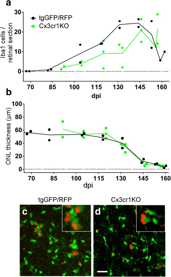
Comparison of microglial migration to photoreceptor area and PR thinning in scrapie-infected mice with and without expression of Cx3cr1. a After 79A scrapie infection, a delay in the number of Iba1-positive cells migrating to the PR areas was seen in Cx3cr1 knockout mice vs tgGFP/RFP (Cx3cr1 heterozygous) mice. Levels became similar in both mouse strains after 145dpi. b No difference was noted in the timing of ONL thinning in Cx3cr1 knockout mice vs tgGFP/RFP mice, suggesting that the delay of microglia had no effect on timing of ONL degeneration. c and d Retinal flat mounts examined by confocal microscopy show green microglia in both tgGFP/RFP mice (c) and Cx3cr1KO mice (d). Both mice also had red/green cells thought to be green microglia which had phagocytosed rhodopsin and/or other outer segment material, which was seen as red autofluorescence [25, 27]. Cx3cr1 mice do not express any RFP, so the red in these mice could not be RFP. Scale bar = 25 μm
