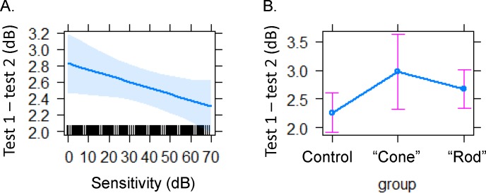
Figure 6.
Variability is lower with higher sensitivity (A) The absolute variability in pointwise sensitivity decreased with higher sensitivities for controls and patients with IRD. (B) Absolute PWS difference between tests was lower for controls and highest for patients in the cone group (autosomal dominant macular, pattern, or cone–rod dystrophy). The rod group (X-linked, X-linked carrier, recessive, isolate, or autosomal dominant retinitis pigmentosa and Usher's syndrome II).

