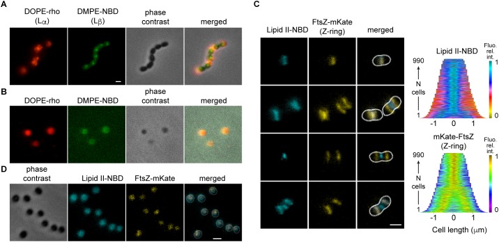
Figure 4.
Localization of lipids in S. pneumoniae depends on the cell shape. (A) Cells were labeled simultaneously with DOPE-rho (red) and DMPE-NBD (green) prior to further growth for 60 min. (B) The cell wall of cells labeled and chased as in (A) was digested by peptidoglycan hydrolases. (C) Growing cells expressing FtsZ-mKate (yellow) labeled with fluorescent lipid II-NBD (cyan) and the corresponding demographs. (D) The cell wall of fixed cells labeled with lipid II-NBD was digested by peptidoglycan hydrolases.

