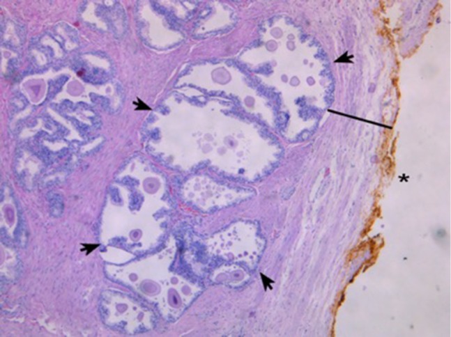
Figure 3.

H&E staining.
Notes: This slide shows an example of a small prostate (24 g) viewed at 50 × magnification. The external, posterior margin is also inked and marked with an asterisk (*). In contrast to Figure 1, a decent number of glands (arrows) are present and easily visible in the peripheral zone (PZ) close to the margin, with a thin capsule (black line) visible.
