
Figure 6.
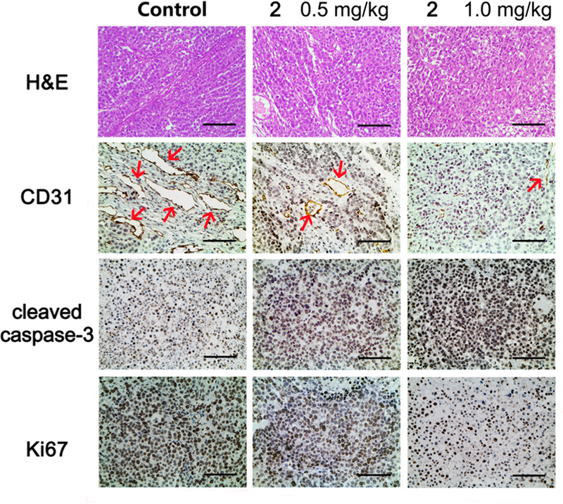
Compound 2 treatment resulted in apoptosis and vascular disruption of NCI-H460 xenograft tumors. Representative images of HE staining and IHC staining of CD31 (endothelial marker), cleaved caspase-3 (apoptosis marker), and Ki67 (proliferation marker) in different treatment groups. Sections were counterstained with hematoxylin. Red arrows in CD31 staining images indicate micro-vessels. Scale bar, 100 μm.