


Figure 2.
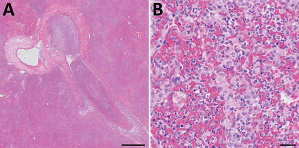
Histopathologic analysis of accessory lung lobe of dog with pneumonic plague (hematoxylin and eosin stain), Colorado, USA. A) Parenchyma, which is diffusely effaced by necrohemorrhagic pneumonia. Scale bar indicates 500 μm. B) Alveolar detail, which is obscured by necrosis, hemorrhage, and suppurative inflammation without intralesional bacteria. Scale bar indicates 20 μm.