
Figure 4.
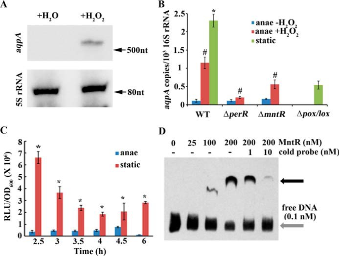
H2O2 induces the expression of the So-aqpA gene. A, anaerobically grown mid–expansion phase cells were collected and divided into two aliquots. One aliquot was treated with 40 μm H2O2 (+H2O2) for 20 min and the other with the same volume of H2O (+H2O) used as a control. Total RNA was extracted from both aliquots, and Northern blotting was performed using a biotin-labeled So-aqpA DNA fragment (Table S1) as the probe. 5S rRNA was used as an internal control (details are described in “Experimental procedures”). B, the tested strains were cultured and treated with 40 μm H2O2 as described in A. Quantitative RT-PCR was performed to quantify the transcript copies of the So-aqpA gene in anaerobically grown (anae −H2O2) and 40 μm H2O2-pulsed (anae +H2O2) wild strain (WT), deletion mutants of perR (ΔperR) and mntR (ΔmntR), and statically grown (static) wild strain and pox and lox double deletion mutant (Δpox/lox) (details are described in “Experimental procedures”). Triplicate measurements were performed for three batches of cultures, and the averages ± S.D. are shown. * and #, data are statistically significantly different in comparison between statically grown WT strain and Δpox/lox mutant (Student's t test, p <0.05) and H2O2 induction on anaerobically grown WT strain and mutants as verified by one-way ANOVA analysis followed by Tukey's post hoc test (p <0.05), respectively. C, a luciferase reporter strain, PaqpA-luc, in which the So-aqpA promoter was fused to the luciferase gene, was grown in BHI broth anaerobically or statically. At the indicated time points during growth, 100 μl cultures were collected in 1.5-ml Eppendorf tubes, and after 5 min exposure to air at room temperature, the luciferase activities (RLU, relative light units) were measured as described in “Experimental procedures.” OD600 was measured in parallel. Triplicate measurements were performed for three batches of cultures, and the averages ± S.D. are shown. *, data are statistically significant compared with those of anaerobically grown WT strain as verified by Student's t test (p <0.05). D, a DNA fragment of the So-aqpA promoter was PCR amplified with 5′-end biotin-labeled primers (Table S1). 0.1 nm biotin-labeled DNA was mixed with various concentrations of MntR protein in the EMSA-binding mixture and run in a native PAGE gel. Black arrow indicates the protein-DNA complex. Addition of increasing nonlabeled DNA (cold probe) decreased the protein-DNA complex.