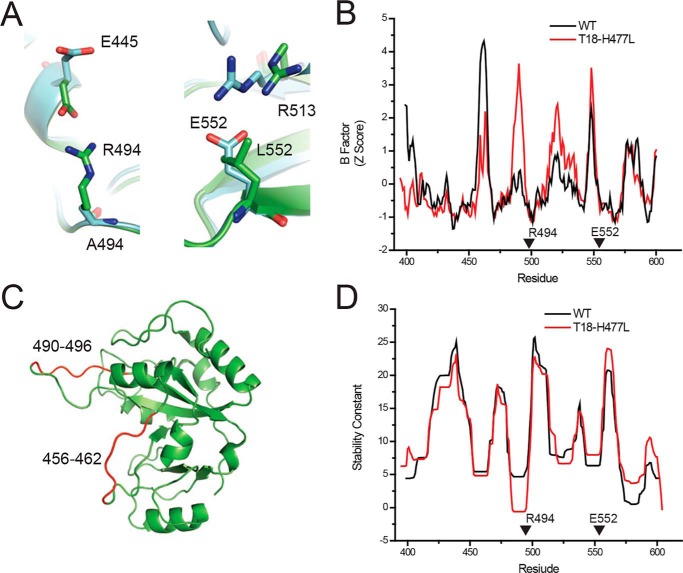
Figure 2.
Deimmunizing mutations alter the structure of PE-III. A, orientation of residues at the 494 and 552 positions in the WT (green, PDB code 1XK9) and T18-H477L (cyan, PDB code 6EDG) structures. B, Comparison of the crystallographic B-factors from the WT and T18-H477L structures. Arrows indicate locations of the R494A and L552E mutations. C, approximate locations of the proteinase K cleavage sites within the WT PE-III structure. D, COREX stability constants of the WT and T18-H477L structures. Arrowheads indicate locations of the R494A and L552E mutations.

