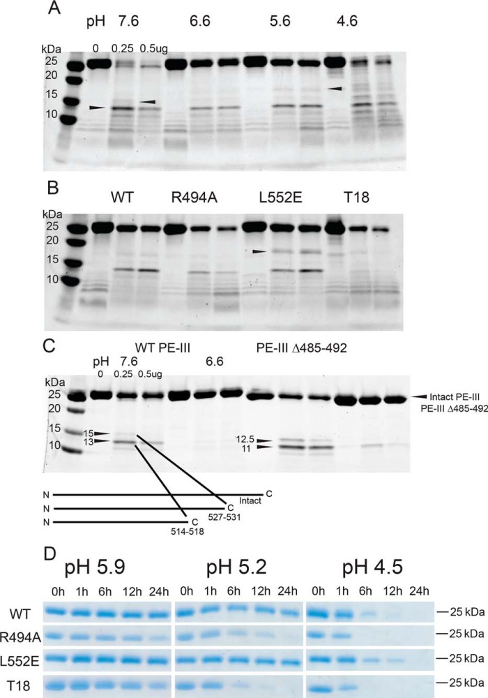
Figure 4.
Destabilized PE-III variants are more susceptible to proteolysis by lysosomal proteases. A, indicated amount of cathepsin S was incubated with WT PE-III for 3 h at 37 °C at the indicated pH prior to analysis by SDS-PAGE and Coomassie staining. Arrowheads indicate fragments selected for analysis by MS. B, limited proteolysis of PE-III variants by cathepsin S at pH 5.6 for 3 h at 37 °C. C, C-terminal cleavage sites were confirmed by limited proteolysis of a PE-III variant lacking the 490s loop mentioned above. Schematic showing the locations of the cathepsin S cleavage sites within the primary structure of PE-III is placed below the gel. D, proteolysis of the PE-III variants using lysosomal extracts at different pH values analyzed by SDS-PAGE and Coomassie staining.

