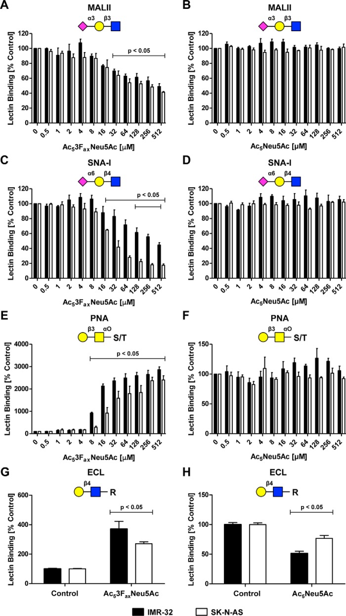
Figure 3.
Effect of sialic acid analogues on overall cell surface sialylation. A–F, IMR-32 cells and SK-N-AS cells were cultured for 3 days with 0–512 μm Ac53FaxNeu5Ac or Ac5Neu5Ac, and cell surface sialylation was assessed by flow cytometry using fluorescent lectins. The lectins recognize α2,3-linked sialic acids (MALII), α2,6-linked sialic acids (SNA-I), or terminal β-galactose (PNA), respectively. Bar diagrams show average binding ± S.E. normalized to control of MALII (A and B), SNA-I (C and D), or PNA (E and F) on IMR-32 cells and SK-N-AS cells treated with Ac53FaxNeu5Ac or Ac5Neu5Ac (n = 3). G–H, IMR-32 and SK-N-AS cells were cultured for 3 days with Ac53FaxNeu5Ac (32 μm) or Ac5Neu5Ac (2 mm). Cells were stained with the ECL lectin, which recognizes uncapped terminal Galβ1–4GlcNAc residues. Bar diagrams show average binding ± S.E. normalized to control of ECL on IMR-32 cells and SK-N-AS cells treated with Ac53FaxNeu5Ac (G) or Ac5Neu5Ac (H) (n = 2).

