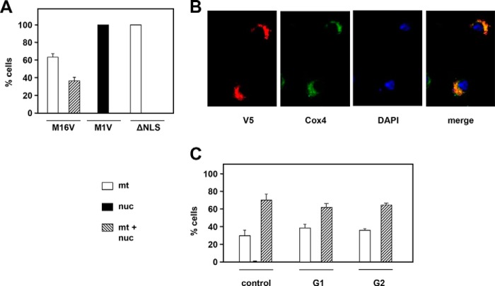
Figure 2.
Subcellular targeting of RNase H1 variants. A, intracellular localization of RNase H1-V5 variants in cultures of stably transfected cells exemplified in B. M1V and M16V, N-terminal methionine variants (see Fig. S2A); ΔNLS, with the putative nuclear localization signal deleted (see Fig. S2C). C, intracellular localization of RNase H1-V5 in cells synchronized in G1 and G2 (see FACS profiles in Fig. S2E). All plotted values are means of three experiments. Error bars denote S.D. nuc, nuclei; mt, mitochondria.

