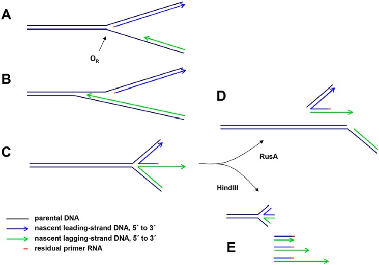
Figure 7.
Proposed interpretations of mtDNA species detected by gel electrophoresis in rnh1 knockdown cells. One of many possible scenarios is illustrated. A, portion of a hypothetical replication intermediate with uncompleted lagging strand approaching the unidirectional replication origin (OR). A short residual RNA primer remains at the 5′ end of the leading strand. B, the lagging strand proceeds beyond the leading-strand initiation point as far as specific, reiterated termination signals in the repeat II elements of the NCR. C, impaired fork progression around the genome causes the origin structure to persist with eventual regression to form a chicken-foot structure that can branch-migrate (arc denoted by blue arrow in Fig. 6C). D, upon treatment with RusA, the four-way junctions resulting from these regressed forks are cut, generating effectively linear products. E, HindIII digestion liberates linear fragments with lagging-strand 3′ ssDNA extensions derived from the regressed forks (green arrows in Fig. 6B). These are digestible with S1 nuclease or exonuclease I, leaving a residual double-stranded species (purple arrow in Fig. 6B).

