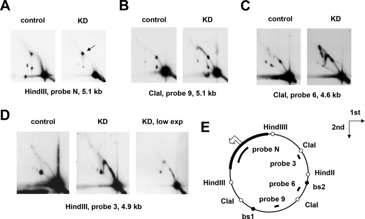
Figure 8.
Knockdown of rnh1 cause accumulation of RIs beyond pause sites. A–D, 2DNAGE of four restriction fragments of Drosophila S2 cell mtDNA, probed as indicated, in material from control cells treated with an inert dsRNA against GFP and cells knocked down for rnh1 by treatment with an rnh1-specific dsRNA (denoted KD). The arrow in A denotes burst bubbles (see text). E, schematic map of Drosophila mtDNA indicating the location of relevant restriction sites (open circles), mTTF-binding sites (bs1 and bs2; filled circles), the noncoding region (bold), and the probes used. The open arrowhead marks the location and direction of replication initiation (see Ref. 40). The directions of the first- and second-dimension electrophoresis in all gels are as indicated by the arrows. The images show relatively low exposures (low exp) to reveal fine details of the arcs of RIs.

