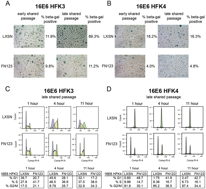
Figure 4. 16E6/FN123 HFKs at shared timepoints had reduced senescent marker expression and maintained cell cycling compared to 16E6/LXSN HFKs.
(A) 16E6/LXSN and 16E6/FN123 HFK3 (A) or HFK4 (B) cells were stained for senescence associated beta-galactosidase (beta-gal) at early and late shared passages (noted as asterisks in Figure 2C and D). (A) At an early shared passage, HFK3 cells were equivalently stained (11.9% vs 9.8%). At a late shared passage, 16E6/LXSN HFK3 had increased staining (89.3% vs 11.2%). (B) At an early shared passage, 16E6/LXSN HFK3 cells had increased staining (18.2% vs 4.0%). At a late shared passage, this was maintained (18.3% vs 4.8%). (C and D) 16E6/LXSN and 16E6/FN123 HFK3 and HFK4 cells were synchronized by density arrest and then released. HFK incorporation of BrdU and propidium iodide were quantified at 1, 4, and 11 hours after release. (C) 16E6/FN123 HFK3 had more than 1½ times as many cells in S phase at 1 hour compared to 16E6/LXSN HFK3, and 16E6/FN123 HFK3 advanced through the cell cycle more rapidly than 16E6/LXSN HFKs at 4 and 11 hours. (D) 16E6/LXSN HFK3 remained in G2/M at 1, 4, and 11 hours after release whereas 16E6/FN123 HFK4 maintained a typical cycling pattern.

