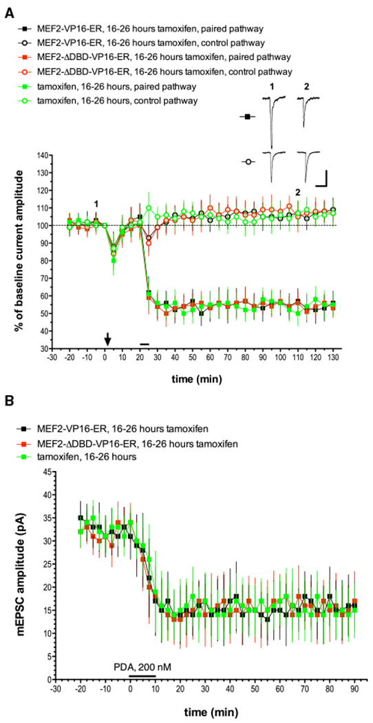
Figure 3. MEF2 Activation Is Not Sufficient to Produce LTD in Purkinje Cells.

Cultured Purkinje cells were transfected with a plasmid that drives expression of tamoxifen-inducible constitutively active MEF2 (MEF2-VP16-ER) or an inactive version that fails to bind DNA (MEF2-VP16-ER-ΔDBD). Wild-type Purkinje cells were transfected with the MEF2-VP16-ER plasmid and then received tamoxifen treatment 3 days later. Recordings were then made 16–26 h after the application of tamoxifen.
(A) Exemplar traces are single (unaveraged) responses, and they correspond to the points indicated on the time course graph. The downward arrow at t = 0 min indicates the delivery of depolarizing pulses alone, without paired glutamate pulses at either pathway. The horizontal bar at t = 20 min indicates glutamate and depolarization pairing delivered to the paired pathway. MEF2-VP16-ER, 16–26 h tamoxifen, n = 8; MEF2-ΔDBD-VP16-ER, 16–26 h tamoxifen, n = 7; tamoxifen alone, 16–26 h, n = 6. Scale bars represent 2 s, 100 pA. When compared with the MEF2-VP16-ER + tamoxifen paired pathway, the late phase of LTD measured at t = 120 min was not significantly different from the paired pathway responses in the MEF2-ΔDBD-VP16-ER + tamoxifen group (p > 0.20) or the tamoxifen-alone control group (p > 0.20).
(B) mEPSC recordings and chemical LTD induction by PDA were performed as indicated for Figure 1B. n = 10 cells/group. When compared with the MEF2-VP16-ER + tamoxifen paired pathway, the late phase of LTD measured at t = 90 min was not significantly different from the responses in the MEF2-ΔDBD-VP16-ER + tamoxifen group (p > 0.20) or the tamoxifen-alone control group (p > 0.20).
