
Fig. 1.
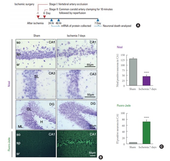
Histological evidence showing 4-vessel occlusion (4-VO) model induces neuronal death in the hippocampal CA1 by 7 days. (A) Time line of ischemic surgery and molecular assay. (B) Representative images of brain sections stained with Nissl and Fluoro-Jade from animals subjected to global ischemia or sham operation 7 days after surgery. (C) Summary data showing the number of living CA1 pyramidal neurons as assessed by Nissl staining (upper), and injured CA1 pyramidal neurons by Fluoro-Jade staining (lower) (n=7 for sham, 4 for ischemic animals). so, stratum oriens; sp, stratum pyramidale; sr, stratum radiatum; ML, molecular layer; GL, granular layer; H, hilus; SL, stratum lacunosum-moleculare. ****P<0.0001. Scale bar is 50 μm.