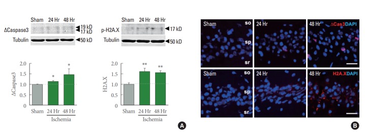
Fig. 2.
Apoptosis and DNA damage induced by ischemic insults. (A) Representative immunoblot images of apoptotic maker, cleavedcaspase-3 and DNA damage marker, phospho-H2A.X. Quantifications were normalized by Tubulin expression (n=7 for sham, 4 for ischemia 24- and 48-hour animals). Values represent mean±standard error of the mean (*P<0.05. **P<0.01). (B) Representative confocal imaging showing, upper, cleaved-caspase-3 (∆Cas3, red) and lower, phospho-H2A.X (red) and counterstained with 4’-6-diamidino-2-phenylindole (DAPI) (blue). Scale bar is 20 µm.

