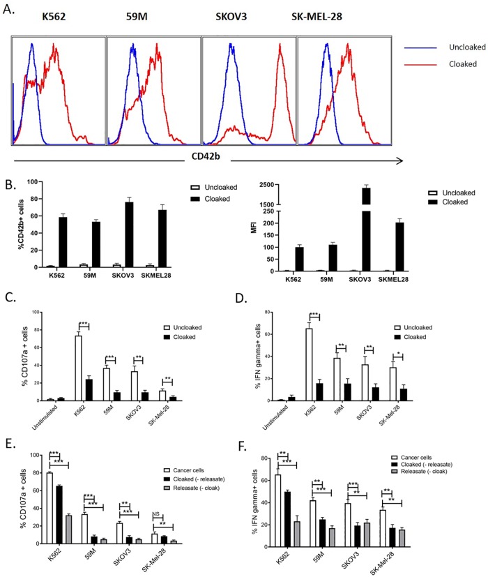
Fig 1. Platelet cloaking inhibits NK cell functions.
(A,B) Quantification of platelet cloaking of ovarian and melanoma tumour cells, and the myelogenous leukaemia control cell line K562. Tumour cell lines were co-incubated with and without platelets and analysed for the surface expression of the CD42b platelet specific marker. Expression data are represented by histogram (A), as a percentage of total cells and by mean fluorescent intensity (MFI; B). (C-F) Analysis of the function consequences of platelet cloaking on NK cell functions. NK cell anti-tumour assays were performed by co-incubating PBMCs with tumour cells (cloaked and uncloaked) for 4 hours and measuring CD107a (C,E) and IFNgamma production (D,F) as markers of activation. (E,F) To dissect the respective roles of platelet contact factors (cloaked (minus the releasate)) and soluble factors (releasate (minus the platelet cloak)) platelets and releasate were isolated and used to treat NK cells in NK activation assays as previously described. (C-F) Data analysed by ANOVA—each experiment represents mean±S.E.M. of at least three independent experiments. * = p<0.05, ** = p<0.01, *** = p<0.001.

