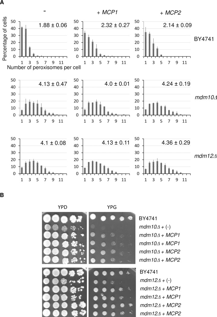
Fig 3. Mcp1 and Mcp2 do not suppress the mdm10Δ and mdm12Δ peroxisome defect.
(A) Percentage of cells for a given number of peroxisomes per cell is shown for the wild type, mdm10Δ and mdm12Δ cells overexpressing either MCP1 or MCP2 or transformed with an empty plasmid (-). Cells were grown on glucose. For each strain, the number of peroxisomes per cell was counted from images of two counts of at least 100 non-budding cells from two independent experiments. Bars represent the SD. On each graph, the average number (± SD) of peroxisomes is indicated. (B) MCP1 and MCP2 restore mdm10Δ and mdm12Δ growth defect on glycerol. Cells of the indicated deletion strains were transformed with a plasmid encoding Mcp1 or Mcp2 or an empty plasmid (-). Growth was analyzed by drop dilution assay on YPD and YPG medium incubated at 28°C for three and five days, respectively. Two different transformants were tested for mdm10Δ and mdm12Δ cells overexpressing the MCP1 or MCP2 gene.

