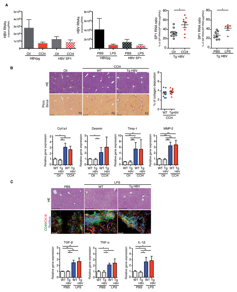
Fig.1. Liver injury increases HBV pgRNA alternate splicing in HBV transgenic mice.
(A) HBV pgRNA and SP1RNA expression (left) and SP1RNA proportion (right) quantified in the liver of TgHBV mice treated with oil/CCl4 for 7 weeks or with PBS/LPS for 2 weeks. (B) Representative H&E and Picro-Sirius red stained histological liver section (left, bar 250μm) and quantification of collagen deposition (right) from 7 weeks oil, CCl4-WT and CCl4-TgHBV treated mice (upper panel). Fibrosis-related mRNA expression in the liver, each dot represents a mouse, experiments were performed on oil-WT (n=7), oil-TgHBV (n=7), CCl4-WT (n=9) and CCl4-TgHBV (n=8) mice (lower panel). C) Representative H&E histological staining (upper, bar: 250μm); CD45 and Cytokeratin 18 (CK18) immunofluorescence staining on liver section (lower, bar: 50μm) from 2 weeks PBS, LPS-WT and LPS-TgHBV treated mice (upper panel). Cytokines mRNA expression in the liver of 2 weeks PBS or LPS-treated WT and TgHBV mice. Each dot represents a mouse; experiments were performed on PBS-WT (n=6), PBS-TgHBV (n=7), LPS-WT (n=5), and LPS-TgHBV (n=5) treated-mice (lower panel). Mann-Withney U-tests: *p<0.05, **p<0.01 and ***p<0.001.

