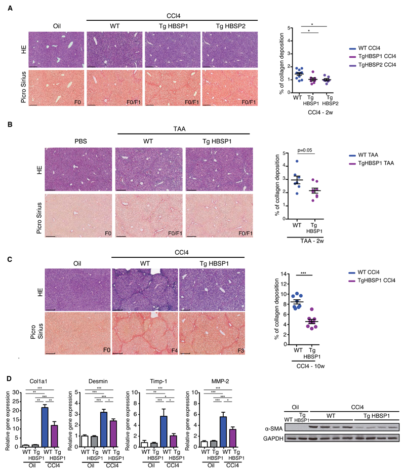
Fig.4. HBSP expression protects against chemical-induced liver fibrosis.
Representative H&E and Picro-Sirius red stained histological liver section (bar 250μm) (left) and quantification of collagen deposition (right) from: (A) WT, TgHBSP1 and TgHBSP2 mice treated for 2 weeks with Oil (n=4), CCl4-WT (n=11), CCl4-TgHBSP1 (n=7) and CCl4-TgHBSP2 (n=7) mice. (B) WT and TgHBSP1 mice treated for 2 weeks with PBS (n=4 for both) or TAA-WT (n=7) and TAA-TgHBSP1 (n=8) mice. (C) WT and TgHBSP1 mice treated for 10 weeks with oil (n=6 for both WT and TgHBSP1) or CCl4 (n=8 for both WT and TgHBSP1). (D) Liver fibrosis-related gene mRNA and α-SMA protein expression in the liver of WT and TgHBSP1 mice treated for 10 weeks with oil or CCl4. Each dot represents a mouse. Mann-Withney U-tests: *p<0.05, **p<0.01 and ***p<0.001.

