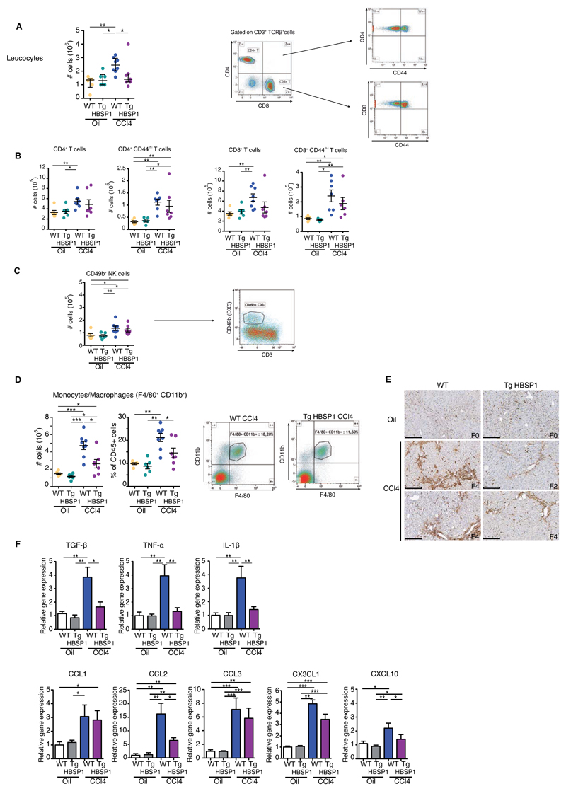
Fig.5. HBSP expression reduces intrahepatic monocytes/macrophages infiltrate.
WT and TgHBSP1 mice were treated for 10 weeks with oil or CCl4. Intrahepatic leucocytes were isolated and analysed by flow cytometry. (A) Intrahepatic leucocyte cells number (left) and representative gating of CD4+ and CD8+ T cells (right). (B) CD4+ T (7-AAD-, CD3+,TCRβ+,CD4+) and CD44hi effector/memory CD4+ T cells number. CD8+ T (7-AAD-, CD3+,TCRβ+,CD8+) and CD44hi effector/memory CD8+ T cells number (left). (C) NK (7-AAD-,CD49b+,CD3-) cells number and representative gating. (D) Intrahepatic monocytes/macrophage cells number, proportion and representative gating of (7-AAD, CD45+, F4/80+, CD11b+) from WT and TgHBSP1 CCl4-treated mice (right). (E) Representative monocytes/macrophages F4/80 staining on histological liver section (bar 250μm). (F) Cytokines (upper panel) and chemokines (lower panel) mRNA expression in the liver. Each dot represents a mouse; experiments performed on 10 weeks oil-WT (n=6), oil-TgHBSP1 (n=6), CCl4-WT (n=8) and CCl4-TgHBSP1 (n=8) treated mice. Mann-Withney U-tests: *p<0.05, **p<0.01 and ***p<0.001.

