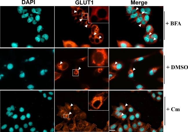
Figure 4.
The localization of GLUT1 in the presence of inhibitors during the chlamydial infection. Hela cell monolayers grown in 8-well chamber slides were infected with C. trachomatis for 6 h, and then incubated with fresh medium containing 1 μg/ml of brefeldin A (BFA), DMSO or 100 μg/ml of chloramphenicol (Cm) for another 12 h. All of the experiments were repeated at least three times with similar results. The arrowheads indicate the chlamydial inclusions. Scale bar, 22.00 μm.

