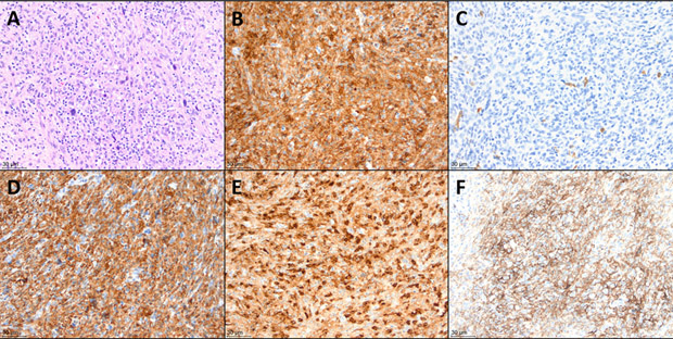
FIGURE 1.
Histologic findings in the patient's initial excisional biopsy (A) Hematoxylin and eosin stain. Atypical histiocytic proliferation with scattered large, hyperchromatic cells, along with background small lymphocytes and occasional eosinophils. (B) CD45 immunohistochemical stain, positive in the atypical cells. (C) CD1a immunohistochemical stain, negative in the atypical cells, while positive in rare background dendritic cells. (D) CD163 stain, positive in the atypical cells. (E) Lysozyme stain, variably positive in the atypical cells. (F) PD-L1 stain, positive in approximately 75% of the atypical cells. All panels are 200× magnification.

