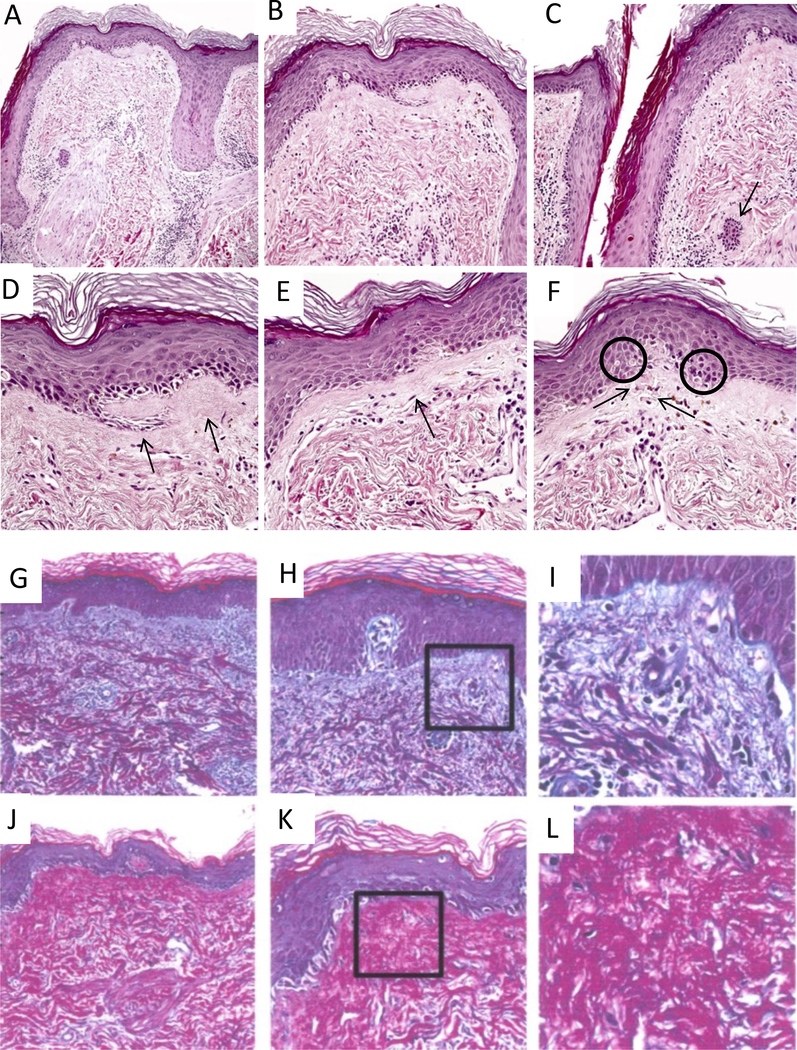
Fig. 2:
Evidence of chronicity in face transplant rejection in this biopsy includes destruction and deformation of hair follicles (f in panel A), patchy perifollicular lymphocytic infiltrates and arrector pili muscles (a, panel A) dissociated from follicles; epidermal thinning with loss of rete ridges (A-F); follicular plugging due to infundibular hyperkeratosis (C); fibrous thickening of subepidermal basement membrane zone (D&E, arrows); and intraepidermal and intradermal colloid body formation (F, encircled and arrows, respectively). Specimen A shows the typical distribution of collagen in null rejection tissue, where type III collagen, shown in blue, resides in the papillary dermis and type I collagen, shown in red, is in the reticular dermis (G-I). Specimen B, however, illustrate the shift of type I collagen into the papillary dermis in the case of chronic rejection (J-L).

