
Figure 4: Splice variants of ADGRG1.
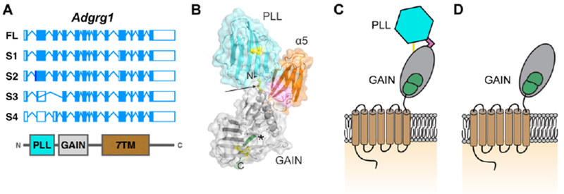
(A) Gene structure of human full-length (FL) ADGRG1 and related isoforms S1-S4. The corresponding protein domains are shown below. Adapted from [32]. (B). Crystal structure of the extracellular domain of ADGRG1 in complex with α5 (orange). Disulfide bonds in yellow, linker in pink. PLL and GAIN domain in cyan and grey, respectively. Modified from [34]. (C). Schematic of ADGRG1 with same color-coding as C. The single yellow line indicating two disulfide bonds between PLL and GAIN domains. (D). Schematic of S4 isoform of ADGRG1.