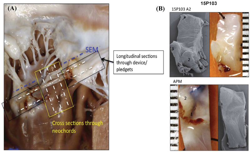
Figure 4:
(A) Schematic of the different cross sections obtained for histopathology and scanning electron microscopy from each sample; (B) Scanning electron microscopy of representative sections from a pig at the leaflet insertion and papillary muscle insertion, which do not depict any thrombus attachment, but demonstrate a small surface that is potentially endothelialized.

