. 2019 Mar 19;60(4):336–345. doi: 10.3349/ymj.2019.60.4.336

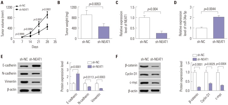
Fig. 5. NEAT1 knockdown represses tumor growth and EMT by Wnt/β-catenin signaling in vivo. Nude mice were subcutaneously injected with 1.0×10⁷ 5-8F cells stably infected by sh-NC or sh-NEAT1, and the mice were euthanized at 5 weeks after implantation. (A) At 7 days after cell implantation, tumor volumes were measured every one week. (B) The average weight of tumors isolated from xenograft mice. qRT-PCR assay of NEAT1 (C) and miR-34a-5p (D) expression levels in excised tumors. Western blot analysis of E-cadherin, N-cadherin, and Vimentin protein levels (E), as well as β-catenin, cyclin D1, and c-myc expression levels (F), in excised tumor tissues with ImageJ software. NEAT1, nuclear paraspeckle assembly transcript 1; EMT, epithelial to mesenchymal transition.

Fig. 5