1.
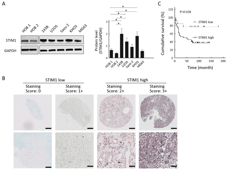
Stromal interaction molecule 1 (STIM1) expression level in osteosarcoma cells and tissue specimens. (A) Western analysis. Cytoplasmic extracts from osteosarcoma cells (143B, U2OS, Saos-2, KHOS and MG63) and human osteoblast (HOB 1 and HOB 2) cells were analyzed by Western blot using anti-STIM1 and anti-glyceraldehyde 3-phosphate dehydrogenase (anti-GAPDH) antibodies as described in methods. Quantitation was performed by densitometry and normalized to GAPDH expression. Data are presented as
 from three independent experiments. *, P<0.05, compared with HOB 1 and HOB 2; (B) Representative immunohistochemical staining of STIM1 in osteosarcoma using tissue microarrays. The lower panel is a magnified view of a region in the respective image in the upper panel. Scale bars are 200 μm in the upper panel and 50 μm in the lower panel; (C) Overall survival of patients in STIM1 high and STIM1 low groups (P=0.028).

