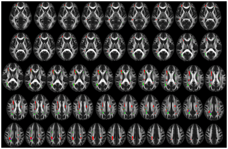
Figure 1. Distinct spatial distribution of white matter hyperintensities related to hypertension and CSF amyloid β1–42 levels.
The spatial distribution of white matter hyperintensities (WMH) related to hypertension (HTN) (red) and Cerebrospinal fluid amyloid beta 1–42 levels (Aβ1–42) (green) shows primarily distinct distributions with minimal overlapping areas (blue). WMH associated with HTN occur primarily in deep cortical white matter and along the body of the lateral ventricles. WMH associated with Aβ1–42 occur primarily near the ventricular horns and the posterior corona radiata. Areas of WMH are displayed on the FMRIB58 FA 1mm3 brain. Contiguous 1mm slices are shown starting from MNI z = 0 at the top left and MNI z = 48 at the bottom right. All images are shown in radiological orientation (anatomical right is on the left side of the image).

