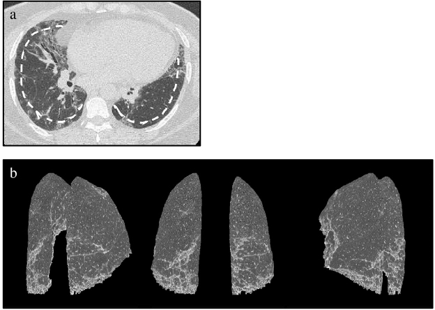
Fig. 2. Patient with representative interstitial lung disease.
(a) Axial HRCT image and dashed line indicating 10-mm depth from the chest wall; (b) multiple-view (posterior view and posterolateral views) 3D-cHRCT images showing the 3D distribution of ILD infiltrates.
3D-cHRCT, three-dimensional curved high-resolution computed tomography; ILD interstitial lung disease

