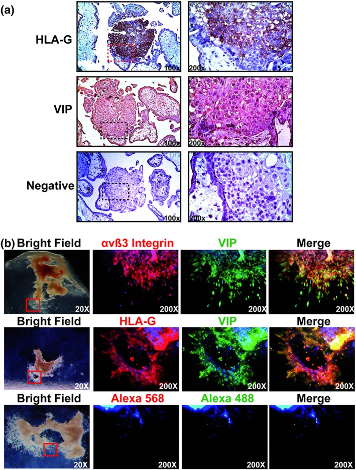
Figure 1.

Columnar cells and EVT cells express VIP. (a) Serial placenta sections were stained with anti‐HLA‐G (1/100) or anti‐VIP (1/500) Abs and haematoxylin. The negative control was incubated with the secondary biotinylated Ab. Microphotographs were taken with an Olympus Microscope with 100× and 200× magnification (squared in the left panel). A representative of 10 different placentas at 5–9 weeks of gestation. (b) Five‐ to 9‐week placenta explants (n = 7) were isolated and plated on a collagen I matrix and cultured with DMEM:F12 10% FCS during 96 hr. The explants were immunostained with anti‐αVβ3 integrin (1/100), anti‐HLA‐G (1/100), or anti‐VIP (1/200) Abs, and the bright field microphotographs were taken with an Olympus microscope whereas the immunofluorescence photographs were taken with a Zeiss Clark microscope with Apotometer. The negative controls incubated with the Alexa's Abs (568 and 488 nm) are shown in the panel above. Ab: antibody; EVT: extravillous trophoblast; FCS: fetal calf serum; HLA‐G: human leukocyte antigen G; VIP: vasoactive intestinal peptide
