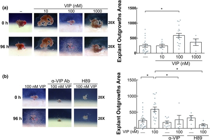
Figure 2.

VIP increases EVT outgrowth in placenta explants through a PKA pathway. (a) Placenta explants were cultured in DMEM:F12 10% FCS without or with VIP (10, 100, and 1,000 nM) during 96 hr. In the left panel, representative microphotographs of the EVT outgrowths are shown. In the right panel, results of the EVT outgrowths area of 15 different placenta explants are expressed as mean ± SEM. Each point depicted represents an individual explant. *P < 0.05. Kruskal–Wallis test and post hoc Dunn's test. (b) The explants were cultured in the absence or presence of 100 nM VIP, neutralizing anti‐VIP (α‐VIP) Ab (1/1000), or H89 (5 μM) similar to panel (a). In the left panel, representative microphotographs are shown, and in the right panel, the results are expressed as mean ± SEM of the EVT outgrowth area of 15 different placenta explants. Kruskal–Wallis test and post hoc Dunn's test. EVT: extravillous trophoblast; FCS: fetal calf serum; VIP: vasoactive intestinal peptide
