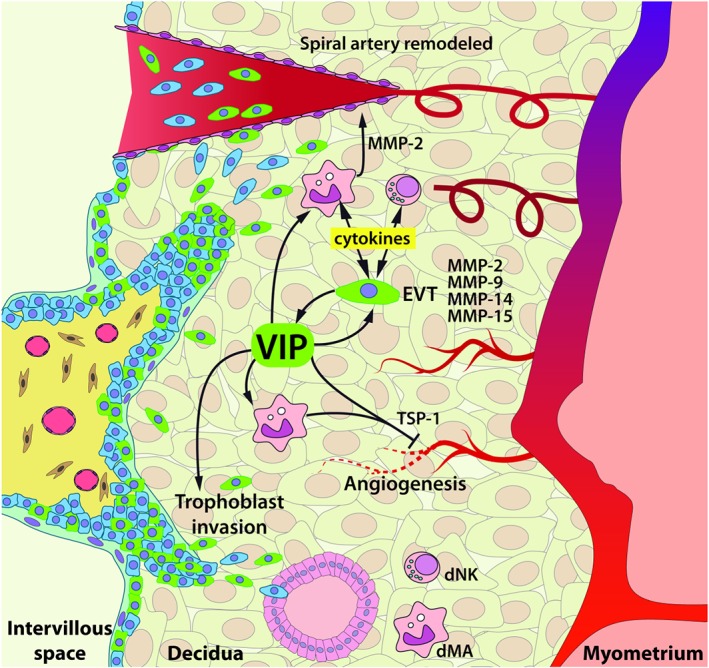
Figure 10.

Hypothetical model of VIP effects in first‐trimester placenta. VIP+ EVT cells (green) contribute to ECM remodelling through MMP‐2, 9, 14, and 15 production and MMP‐2 by dMA primed with VIP. Along with this, the immune micro‐environment is shaped to a “silent” milieu with an upregulation of IL‐10 and chemokine production and decrease or no changes in pro‐inflammatory cytokines by EVT, dNK cells, and dMA. VIP+ EVT cells invade the decidual stroma, transform maternal spiral arteries, and are lining the glandular epithelium. VIP regulates EC tube formation directly or indirectly through dMA with high expression of TSP‐1. dMA: decidual macrophages; dNK: decidual natural killer; EC: endothelial cells; ECM: extracellular matrix; EVT: extravillous trophoblast; MMP: metalloproteinase; TSP‐1: thrombospondin‐1; VIP: vasoactive intestinal peptide
