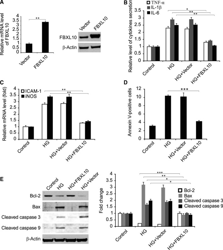
Figure 2.

FBXL10 reduced HG‐induced inflammatory response and apoptosis. (A) H9c2 cells were transfected with FBXL10, FBXL10 expression was analyzed by Western blotting and real‐time RT‐PCR. Data represent the mean ± SD of three independent experiments. **P < 0.01. (B) H9c2 cells with or without FBXL10 transfection were stimulated by HG. The secretion of TNF‐α, IL‐1β and IL‐6 was determined by ELISA. Data represent the mean ± SD of three independent experiments. **P < 0.01; *P < 0.05. (C) H9c2 cells with or without FBXL10 transfection were stimulated by HG. The expression of ICAM‐1 and iNOS was assessed by real time RT‐PCR. Data represent the mean ± SD of three independent experiments. **P < 0.01. (D) H9c2 cells with or without FBXL10 transfection were stimulated by HG. Cell apoptosis was assessed by flow cytometry. Data represent the mean ± SD of three independent experiments. ***P < 0.001. (E) H9c2 cells with or without FBXL10 transfection were stimulated by HG. Indicated protein level was detected by Western blotting and normalized to β‐actin. Data represent the mean ± SD of three independent experiments. ***P < 0.001; **P < 0.01; *P < 0.05
