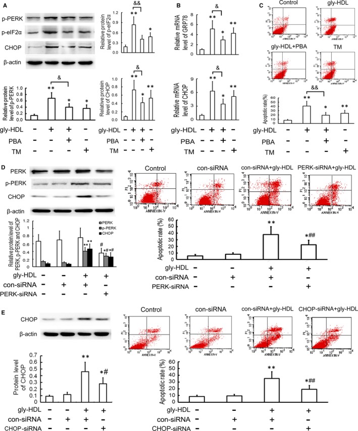
Figure 3.

Attenuation of ER stress‐CHOP pathway inhibits gly‐HDL‐induced macrophage apoptosis. A and B, RAW264.7 cells were exposed to 100 mg/L gly‐HDL or TM (4 mg/L) in the presence or absence of PBA (5 mmol/L) for 24 h, and then the protein and mRNA levels of ER stress markers were measured by Western blotting and quantitative real‐time PCR, respectively. C, Cell apoptosis was determined by flow cytometry and the total apoptotic cells were shown on the right side of the panel (Annexin V staining alone or together with PI). D and E, RAW264.7 cells were transfected with siRNA specific for PERK or CHOP, treated with 100 mg/L gly‐HDL for 24 h, and then PERK, p‐PERK and CHOP protein levels and cell apoptosis were analysed by Western blotting and flow cytometry, respectively. Data are expressed as the mean ± SD of at least three independent experiments. *P < 0.05, **P < 0.01 vs control group; & P < 0.05, && P < 0.01; # P < 0.05, ## P < 0.01 vs gly‐HDL group transfected with con‐siRNA
