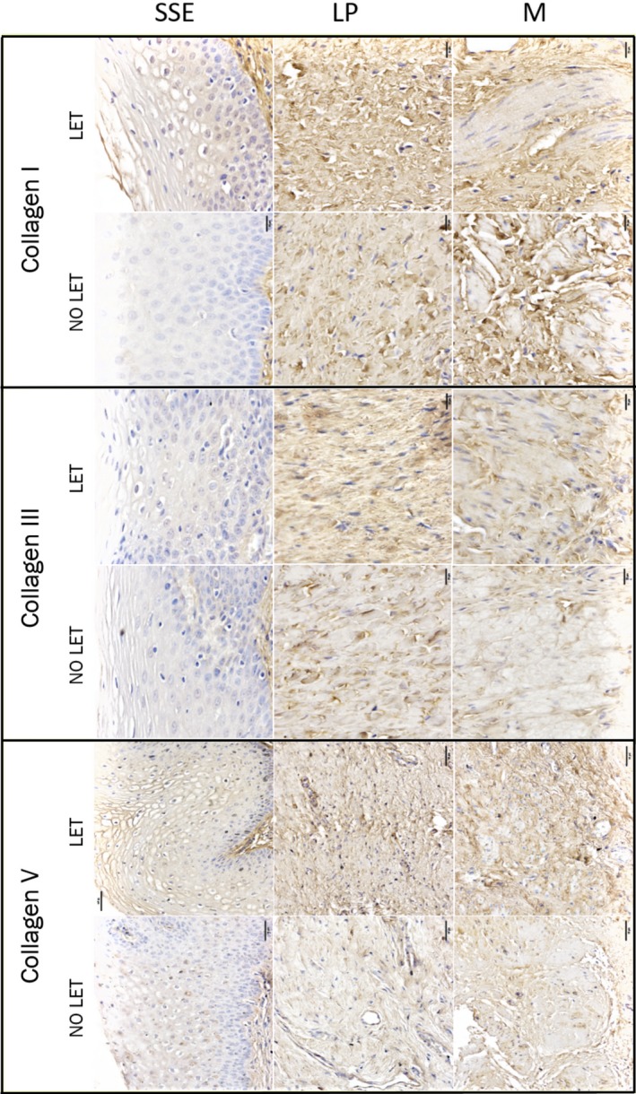
Figure 2.

Immunohistochemical localization of Collagen I, III, and V within human vaginal biopsy samples of post‐menopausal women with severe POP. Shown are representative images of vaginal tissues from POP patients treated with LET and no‐treated controls. The immunolabelling for collagen proteins is indicated by brown deposit. Magnification is 200×; scale bar 50 μm. Negative control is shown on Figure S1
