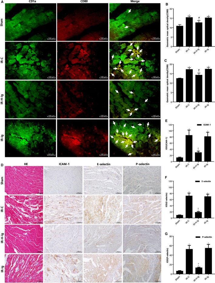
Figure 2.

HMGB1‐TLR4 signalling pathway mediates the migration, adhesion and activation of dendritic cells (DCs) in ischaemia‐reperfusion myocardium. A, Double immunofluorescence of DCs in myocardial tissue among all groups (n = 10 for each group). Samples were collected right after IR procedure and staining was performed as described in Section 2. Representative fluorescence images (200×) of the distribution of CD1a (green), of CD80 (red). Arrows indicate co‐localization. (B,C) The geometric mean optical density of CD80, CD86 among all groups (n = 10 for each group) in peripheral blood of rats was detected by flow cytometry. Histological study, immunohistochemical staining in cardiac tissues from rats of different groups (n = 10 for each group). Samples were collected right after IR procedure was over and staining. D, Hematoxylin‐eosin (HE) staining pictures (200×) are shown in the left, immunohistochemical staining pictures (200×) are in the middle and right; (E) integral optical density (IOD) of ICAM‐1; (F) IOD of E‐selectin; (G) IOD of P‐selectin. Scale bars, 100 μm. *P < 0.05, comparisons of IR‐C, IR‐H‐Ig and IR‐Ig groups with Sham group; #P < 0.05, comparisons of IR‐H‐Ig and IR‐Ig group with IR‐C group
