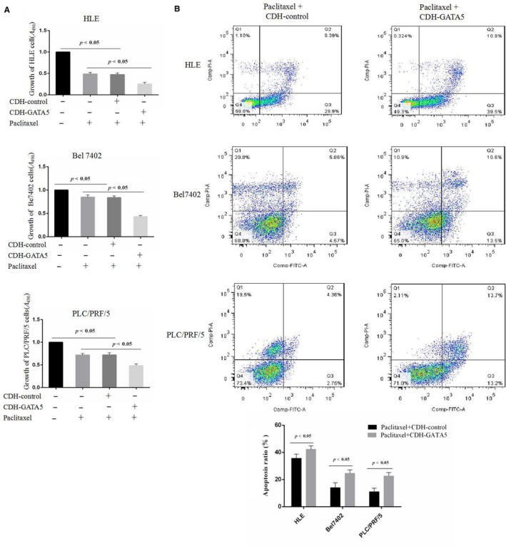
Figure 5.

Influence of GATA5 and paclitaxel on the growth and apoptosis in HCC cells. A, HLE, Bel 7402 and PLC/PRF/5 cells were transfected with CDH empty vector and CDH‐GATA5 followed by treatment with paclitaxel (20 μg/mL) for 24 h. HCC growth was assessed by MTT P < 0.05 indicates statistical significance, N = 6. B, HLE, Bel 7402 and PLC/PRF/5 cells were transfected with CDH empty vector and CDH‐GATA5 followed by treatment with paclitaxel (20 μg/mL) for 24 h. Apoptosis in HLE cells was assessed by flow cytometry. The low columnar picture is the statistical analysis of the apoptosis ratios. P < 0.05 indicates statistical significance
