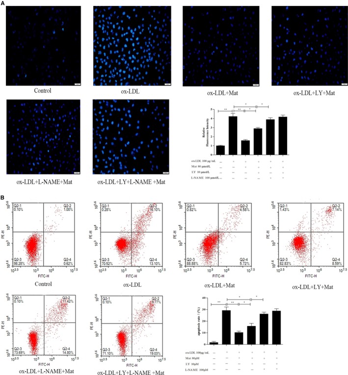
Figure 4.

Effects of matrine on ox‐LDL‐induced apoptosis in HUVECs. A, Hoechst 33258 staining in cultured HUVECs, fluorescence intensities were measured using a fluorescence microscope; representative fluorescence images are shown (100×). B, Apoptosis analysed using Annexin V‐FITC/PI staining in a flow cytometry assay. Representative images of cell populations are shown and quantitative assessment of three independent cell apoptosis experiments was performed. Three independent samples were used. Data are shown as the mean ± SD, *P < 0.05, **P < 0.01
