Figure 1.

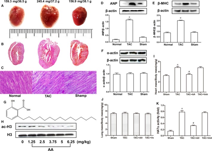
Anacardic acid (AA) blunts pressure overload‐induced hypertrophy in mouse hearts. Stereoscope and haematoxylin and eosin staining analyses of the mouse hearts treated by thoracic aortic banding (TAB) showed that they were significantly inflated compared with those of the sham group, but myocardial hypertrophy, particularly in the left ventricular and interventricular septum, was observed in the transverse aortic constriction (TAC) mice. (A) Characteristic images of complete mouse hearts. (B) Haematoxylin and eosin‐stained longitudinal sections. (C) Longitudinal view of cardiomyocytes. Scale = 20 μm in C. Additionally, the myocardial hypertrophy biomarkers atrial natriuretic peptide (ANP) and myosin heavy chain (β‐MHC) were increased significantly in the TAC mouse hearts compared with those of the sham group (D and E), but the expression levels of α‐actin remained the same in similar samples (F). (G) The chemical structure of the histone acetylase (HAT) inhibitor AA. (H) Different concentrations of AA were used to determine the optimal exposure dose of AA, and 3.75 mg/kg was selected based on the ac‐H3 level. (I‐J) Quantitative analysis of cardiac mass index and lung mass index. (K) The HAT activity was increased significantly in the TAC mouse hearts compared to that of the sham group, but HAT activity was reduced in the hearts of mice treated with AA. *P < 0.05 vs the sham group, # P < 0.05 vs TAC (n = 6)
