
Figure 2.
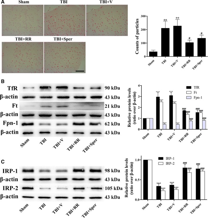
TBI induced cellular iron accumulation while ruthenium red (RR) or Sper administration partially reversed the effects. (A) Mice were subjected to traumatic brain injury (TBI) and then administrated RR (3 mg/kg), Sper (5 mg/kg) or vehicle 30 min after TBI. Ipsilateral cortex was collected 1 day after TBI and then subjected to iron staining. The left panel shows representative brain sections of cortex with iron stained (red) cells. The right panel is quantification of the digitized images showing the count of particles and fraction of total iron staining in each group. (B, C) TBI‐induced disruption of cellular iron homeostasis was improved by RR or Sper. Mice were subjected to TBI and treated with RR (3 mg/kg), Sper (5 mg/kg) or vehicle 30 min after TBI. Ipsilateral brain tissues were collected 1 day after TBI. The protein levels of TfR, Ft, Fpn‐1, IRP‐1 and IRP‐2 were measured by Western blot. TfR and Ft proteins were up‐regulated while Fpn‐1, IRP‐1 and IRP‐2 proteins were down‐regulated after TBI, however RR or Sper treatment reversed these effects. Data are presented as mean ± SEM, n = 6 per group; **P < 0.01, ***P < 0.001 vs sham group; # P < 0.05, ### P < 0.001 vs TBI + vehicle group. β‐actin was used as a loading control