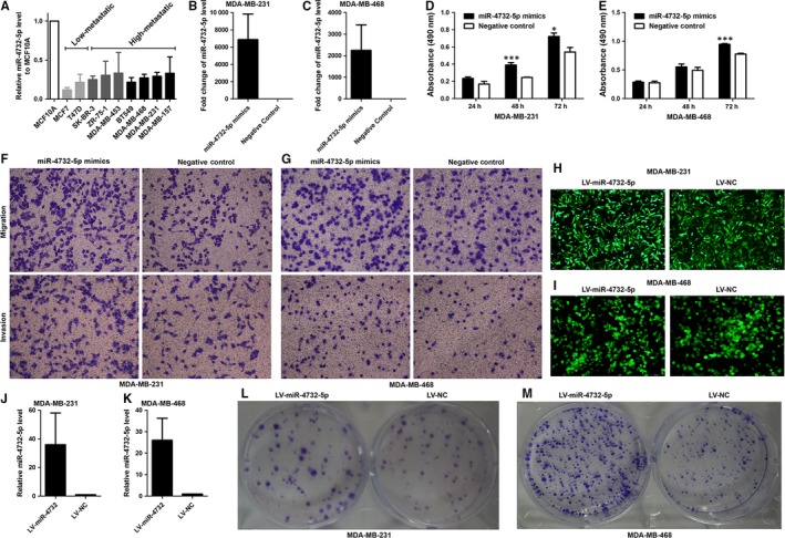
Figure 2.

Expression of miR‐4732‐5p in breast cancer cell lines and its effect on cell biological behaviours. (A) miR‐4732‐5p was down‐regulated in breast cancer cell lines (n = 9) compared with the non‐tumourigenic cell line MCF10A. It is also noted that miR‐4732‐5p was relatively highly expressed in high‐metastatic cell lines than low‐metastatic cell lines. (B‐C) miR‐4732‐5p mimics transfection led to significant high expression of miR‐4732‐5p in breast cancer cells. (D‐E) Overexpression of miR‐4732‐5p promoted cell proliferation as revealed by MTS assays. (F‐G) MiR‐4732‐5p enhanced cell migration and invasion ability, compared with negative control. (H‐I) After lentivirus vector transfection, green fluorescence protein expression was observed by using fluorescence microscope. (J‐K) Lentivirus miR‐4732‐5p vector up‐regulated miR‐4732‐5p expression, compared with the control vector. (L‐M) Stable expression of miR‐4732‐5p expression increased colony formation in MDA‐MB‐231 and MDA‐MB‐468 cells. *P < 0.05; **P < 0.01
