
Figure 4.
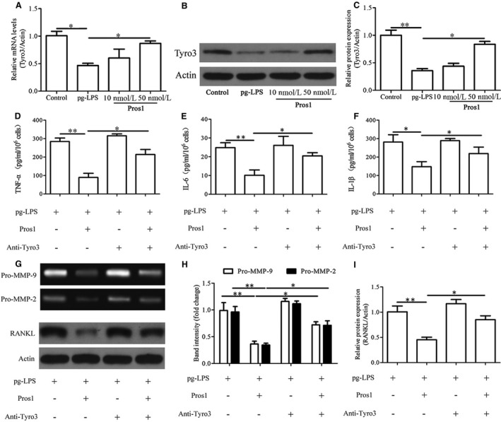
Pros1 inhibits p.g‐LPS–induced inflammation in hGECs via Tyro3. (A‐C) hGECs were treated with p.g‐LPS (1 μg/mL) and Pros1 (10 or 50 nmol/L), alone or in combination as indicated for 24 hours. Untreated cells (Control) served as control. Tyro3 mRNA (A) and protein (B, C) levels were determined by qRT‐PCR and Western blot analysis respectively. (D‐I) hGECs were treated with p.g‐LPS (1 μg/mL), Pros1 (50 nmol/L) and anti‐Tyro3 antibody (10 μg/mL), alone or in combination as indicated for 24 hours. (D‐F) TNF‐α (D), IL‐6 (E) and IL‐1β (F) protein concentrations in the culture supernatants were determined by ELISA. (G, H) The enzymatic activities of MMP‐9 and MMP‐2 in the culture supernatants were assessed by gelatin zymography. (G, I) RANKL protein levels were determined by Western blot analysis. n = 3, *P < 0.05, **P < 0.01