
Figure 1.
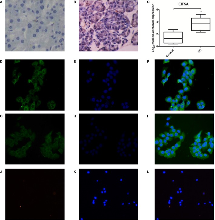
Staining of EIF5A in human PC tissue sections and PC cells. A, Immunohistochemistry staining of EIF5A in human normal pancreatic tissue sections (n = 5). B, Immunohistochemistry staining of EIF5A in human PC tissue sections (n = 30). C, Graphs showing quantitative analyses of EIF5A levels in PC patient samples. D‐I, Immunofluorescence staining of EIF5A in the Panc‐1 and BxPc‐3 cell lines. J‐L, Immunofluorescence staining of EIF5A in pancreatic stellate cells. *P < 0.05, compared with the control (normal pancreatic tissue), as determined by the Student's t test