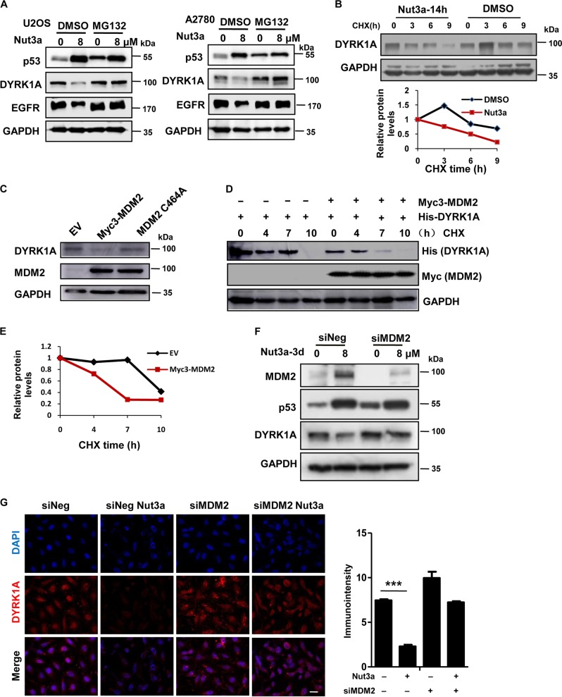
Fig. 5. Negative regulation of DYRK1A by p53 is mediated by MDM2.
a Downregulation of dual-specificity tyrosine-phosphorylated and tyrosine-regulated kinase 1A (DYRK1A) by p53 activation is attenuated by MG132. U2OS and A2780 cells were treated with 8 μM Nut3a for 48 h, and were then treated with 10 μM MG132 for the last 6 h before harvest. Protein levels of p53, epidermal growth factor receptor (EGFR), and DYRK1A were analyzed by western blot. b Nut3a shortens the half-life of DYRK1A proteins. U2OS cells were treated with dimethyl sulfoxide (DMSO) or 8 μM Nut3a for 17 h, and were then treated with 50 μg/mL cycloheximide for the indicated durations before harvest. Protein levels of DYRK1A were analyzed by western blot. Bottom: Signals on the immunoblots were analyzed by the ImageJ software (NIH, Bethesda, MD, USA)45 and the DYRK1A protein amounts were normalized with that of glyceraldehyde 3-phosphate dehydrogenase (GAPDH). c Ectopic expression of MDM2 downregulates DYRK1A. pCMV-myc3-HDM2 (WT) and MDM2 C464A expression vectors were respectively transfected into U2OS cells using Lipofectamine 2000, and cells transfected with empty vector were as control. After 48 h, the cells extracts were examined by western blot for the determination of MDM2, DYRK1A. d MDM2 shortens the half-life of DYRK1A proteins. Co-transfection of His-tagged DYRK1A and Myc-tagged MDM2 or empty vector into U2OS cells, and cells were then treated with 50 μg/mL cycloheximide for the indicated durations before harvest. Protein levels of DYRK1A and MDM2 were analyzed by western blot. e Signals on the immunoblots of Fig. 5d were analyzed by the ImageJ software (NIH, Bethesda, MD, USA)44 and the DYRK1A protein amounts were normalized with that of GAPDH. f Downregulation of DYRK1A by p53 activation requires MDM2. U2OS cells were transfected with small interfering RNA (siRNA) duplexes (200 nM) specific to MDM2 or negative oligo in serum-free medium for 4 h, and then were incubated with complete medium for 48 h. The protein levels of MDM2, p53, and DYRK1A in U2OS (siNeg and siMDM2) were measured 72 h after treatment with Nut3a. g Downregulation of DYRK1A by p53 activation requires MDM2, as determined by immunofluorescence. Scale bar, 10 μm. Right, quantitative immunointensity of DYRK1A was analyzed by the ImageJ software (NIH, Bethesda, MD, USA) *** p < 0.001 vs. control

