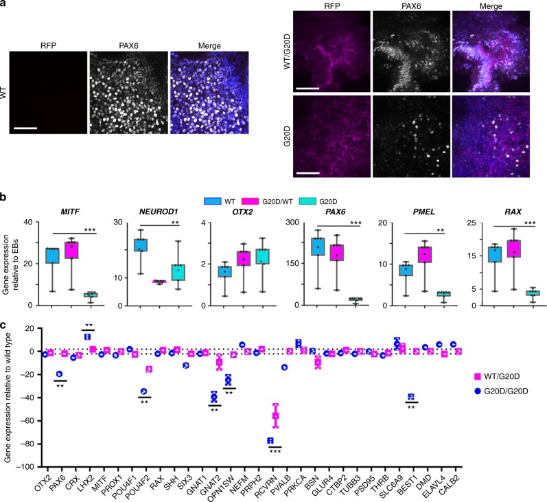
Fig. 3.
CLDN19G20D,G20D down-regulated the expression of markers for retinal development. Clones were analyzed on 30 days of differentiation (DD30) (a, b) or DD80 (c). a Confocal imaging. Optic vesicles expressed PAX6 in CLDN19WT,WT and CLDN19WT,G20D clones but not CLDN19G20D,G20D. Puromycin-red fluorescent protein (RFP) was evident in the transduced clones. b Expression of messenger RNAs (mRNAs) was determined by quantitative-real-time RT-PCR (qRT2-PCR), normalized to glyceraldehyde 3-phosphate dehydrogenase (GAPDH) and compared to expression in undifferentiated embryoid bodies (EBs). c Expression of mRNAs was normalized to GAPDH and compared to expression in clones of CLDN19WT,WT. Scale bar, 20 µm; error bars: SEM for three isolates each of two (G20D) or three (G20D/WT) clones. **P < 0.01, ***P < 0.001

