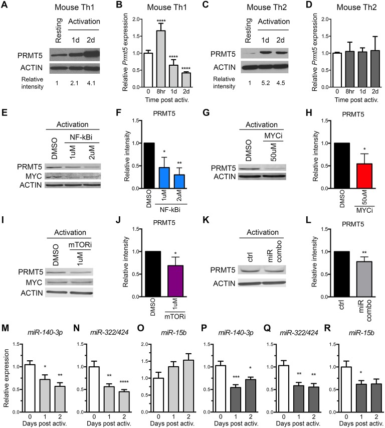
Figure 3.
Prmt5-targeting miRNAs are downregulated after memory murine Th cell activation. (A–D) Murine memory MBP TcR transgenic Th1 (A,B) or Th2 (C,D) line cells (10) were activated with anti-CD3/CD28 and PRMT5 protein expression (A,C, western blot) and Prmt5 mRNA transcripts (B,D, real-time PCR) were analyzed at the indicated time-points. Resting cells are Th1/Th2 line cells 7 days after restimulation with antigen presenting cells and MBP. Data are pooled from 3 to 4 independent experiments. (E–J) Memory mTh1 cells were activated and treated with NF-κB inhibitor Bay11 (NF-κBi), MYC inhibitor 10058-F4 (MYCi), mTOR inhibitor rapamycin (mTORi), or DMSO vehicle control. PRMT5 and MYC protein expression were measured by Western blot (E,G,I) and quantified by ImageStudio (F,H,J). ACTIN is used as a housekeeping control. (K,L) Memory Th cells were activated and transfected with a combination of miR-15b, miR-140-3p and miR322/424. PRMT5 protein expression was analyzed by western blot t and quantified by ImageStudio (L), n = 5. Data are pooled from three independent experiments. Experiments in which transfection efficiency was lower than 20% were excluded from analysis. miR-140-3p, (M,P) miR-322/424 (N,Q), and miR-15b (O,R) expression was analyzed by Real Time PCR in Th1 (M–O, light gray bars) and Th2 (P–R, dark gray bars) and expressed as fold change relative to resting baseline. Data are pooled from four independent experiments (n = 8). *p < 0.05, **p < 0.01, ***p < 0.001, ****p < 0.0001, one-way ANOVA, followed by Dunnett's multiple correction test.

